Can Skin Grafts Be Rejected? Will a Skin Graft for a Foot Wound Take?
A graft of your own skin raises no immune matching problem; when it fails, the cause is fluid, rubbing, infection or blood flow. How a graft takes, heals and lasts on the foot.
Medically reviewed by Dr. Efe Ozturk · Last reviewed
Published 7 min read
Two worries come up first: will it work, and will my body fight it? Here is how a skin graft for a foot wound attaches, why your own skin isn’t rejected, what makes a graft fail and what you’ll see as it heals. For who the operation suits and how it is done, see skin grafts and flaps.
Why a skin graft is often the faster way to close a foot wound
When a clean wound is too large to close on its own, a graft can cover it in one operation. In a study of 100 people with diabetic foot ulcers, the 50 who chose a skin graft healed significantly faster and left the hospital sooner than the 50 treated with dressings; every grafted ulcer healed.[6] The authors of the 11-study review concluded that grafting should be used more often for clean, hard-to-heal ulcers of the leg and the top of the foot.[2]
Dr. Efe Ozturk, a Foot and Ankle Surgeon who is double board certified, including in wound care (FAPWH), performs split-thickness skin grafts for the foot wounds that need them, once infection is cleared and blood flow has been checked.
Can skin grafts be rejected?
It depends on where the skin comes from.[1]
- Your own skin (an autograft). Taken from your own body, it raises no immune matching concerns. It survives as tiny new blood vessels grow into it from the wound bed.
- Donor skin (an allograft). Skin from a deceased donor works as a temporary living dressing. It carries risks of rejection and disease transmission.
- Animal skin (a xenograft). Pig skin is used for temporary coverage; it does not grow a new blood supply.
In burn care, donor skin covers wounds until your own skin can be grafted.[7] Lab-made and donor-derived wound products are a separate group; see skin substitutes. When a graft of your own skin doesn’t take, the cause is not rejection.
How do skin grafts work? The healing stages
The harvest. A split-thickness graft takes the outer layer of skin and part of the layer beneath it, often from the outer thigh. It can be meshed with small slits so fluid drains out from under it, which lowers the risk of a blood or fluid pocket. Meshed grafts are more fragile and take longer to fill in than unmeshed ones.[4]
The first two days. A graft has no blood supply of its own.[4] For about 48 hours, it “drinks” nutrients and oxygen from fluid seeping up from the wound bed, a stage called plasmatic imbibition.[8]
Plugging in. Next, new vessels growing up from the wound bed connect with the graft’s own vessels (inosculation), which brings blood flow back quickly.[8]
The weeks after. In one study, the whole graft had taken in 84% of people at day five and in 62% at weeks three and eight, yet every grafted wound went on to heal completely.[6] The donor site is a shallow wound that resurfaces within two to three weeks.[4]
The months after. All grafts shrink a little over time. A healed split-thickness graft looks smoother and paler, can change color, and a meshed graft has a different texture, less feeling and is more easily injured.[4]
Will my skin graft take? What the results show
Across 11 studies, 85.5% of grafted ulcers healed in about 5.4 weeks, and 12.1% needed regrafting.[2] In two single-center series, 28% and 19.1% of people needed a second graft or a revision, and in the series of 83 people with diabetes, every wound was closed by final follow-up.[9,10] A second graft is a normal part of this treatment.
What makes a skin graft fail?
- Fluid, blood and rubbing. Swelling fluid, drainage or a blood pocket under the graft can lift it off the wound, as can shearing (sliding) forces.[3]
- Infection. Infection affected 4.4% of grafted ulcers in the pooled studies.[2] In a laboratory model of a wound, staph and strep bacteria, helped by plasminogen (a protein in blood), broke down the fibrin “glue” that holds a fresh graft to its bed.[11] A clean, infection-free wound bed improves take.[4]
- Blood flow. Because the graft lives on the wound bed, that bed needs a good blood supply.[4] We check circulation at our practice and, if it is reduced, refer you to a vascular specialist before grafting; see circulation tests for foot wounds.
- Your health. A current or past smoking habit was linked to graft complications.[9] People on dialysis needed revision about three times as often (46.2% versus 14.8%).[10] In one study, people with sleep apnea had graft failure in 30.6% of wounds, against 15.9% without it.[12] Read why smoking and diabetes slow surgical healing (external site).
How a wound VAC and offloading protect the graft
A wound VAC (negative-pressure wound therapy) can be placed over a fresh graft as its holding dressing. Across 16 randomized trials of skin grafts, graft take was 8.3% higher than with standard dressings, the odds of losing the graft were more than halved, and the odds of another operation were lower.[5] It helps by preventing lift-off and reducing shear.[3] In 11 foot and ankle wounds in people with diabetes, grafts held this way healed in 17 days on average.[13] We use negative-pressure wound therapy for the wounds that need it.
Pressure matters too. On the sole, careful patient selection and offloading after surgery helped grafts hold up over the long term.[14] For a graft on the bottom of the foot, we fit an offloading device and show you how to get around safely.
Can skin grafts fail years later?
Once your own skin has taken, it is living skin. The later risk is a new sore in the same place. In the pooled studies, 4.2% of healed ulcers came back over about two years.[2] In a study of 182 people, wounds came back in 17% of grafts on the sole and 10% elsewhere, not a significant difference.[14] If the pressure that caused the ulcer remains, surgery that takes pressure off can correct the bone or tendon behind it.
Limits of the evidence
Most foot graft studies look back at one center’s patients. Grafts on the sole were less often healed at one year (38% versus 64% elsewhere), though time to full healing was similar.[14] Donor-site infection occurred in about 1.7%.[2] In the high-risk series, 5 of the 18 people who needed a revision later had a major amputation.[10] The wound VAC trials covered grafts anywhere on the body, and the results varied between trials.[5]
Common questions
Can skin grafts be rejected?
A graft of your own skin raises no immune matching problem. Donor skin can be rejected and is used as a temporary cover.[1]
Can skin grafts fail years later?
The graft itself becomes your skin, but a new ulcer can form on it if pressure continues. Over about two years, 4.2% of grafted ulcers returned in pooled studies.[2]
Which skin graft is right for an open wound?
For a clean wound with good blood flow, your own split-thickness skin is the usual choice. Thicker grafts suit areas under heavy stress, such as the sole, but need a richer blood supply and are less suited to long-standing wounds.[4] Donor or animal skin can serve as a temporary cover first.[1]
What are the stages of skin graft healing?
For about two days the graft feeds on wound fluid, then new vessels connect to it.[8] The donor site resurfaces in two to three weeks, and the graft keeps maturing and shrinking slightly over time.[4]
The bottom line
A graft of your own skin is not rejected, and it heals most clean foot wounds; when part doesn’t take, a second graft can finish the job. Clearing infection, confirming blood flow, holding the graft still and keeping weight off it give it the strongest start. If a clean foot wound has stalled, ask whether a graft can close it sooner.
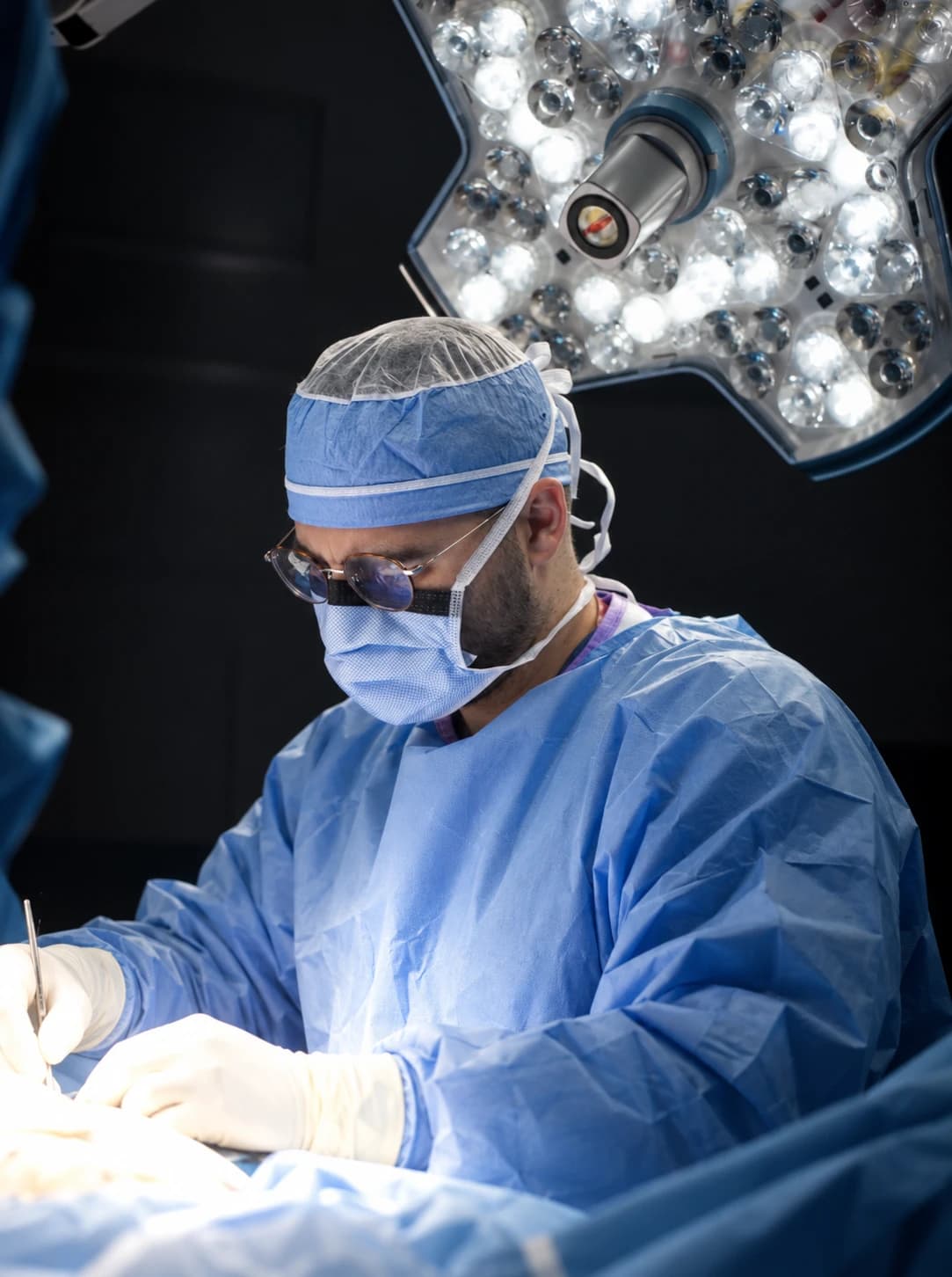
Reviewing surgeon
Foot and Ankle Surgeon at Limb Salvage Institute, a specialty practice of Ozturk Foot & Ankle. Dr. Ozturk is double board certified, as a Diplomate of the American Board of Podiatric Medicine (DABPM) and a Fellow of the Academy of Physicians in Wound Healing (FAPWH), and is a Fellow of the American Society of Podiatric Surgeons (FASPS) and the American Society of Podiatric Medicine (FASPM).
References
- 1.Elseth A, Nunez Lopez O. Wound Grafts. In: StatPearls. Treasure Island (FL): StatPearls Publishing; 2025. PubMed 33232052 (external site)
- 2.Yammine K, Assi C. A Meta-Analysis of the Outcomes of Split-Thickness Skin Graft on Diabetic Leg and Foot Ulcers. Int J Low Extrem Wounds. 2019;18(1):23-30. PubMed 30841767 (external site)
- 3.Azzopardi EA, Boyce DE, Dickson WA, et al. Application of topical negative pressure (vacuum-assisted closure) to split-thickness skin grafts: a structured evidence-based review. Ann Plast Surg. 2013;70(1):23-9. PubMed 23249474 (external site)
- 4.Braza ME, Marietta M, Fahrenkopf MP. Split-Thickness Skin Grafts. In: StatPearls. Treasure Island (FL): StatPearls Publishing; 2025. PubMed 31855388 (external site)
- 5.Lee SC, Bayan L, Sato A, et al. Benefits of negative pressure wound therapy in skin grafts: A systematic review and meta-analysis of randomised controlled trials. J Plast Reconstr Aesthet Surg. 2025;102:204-217. PubMed 39932531 (external site)
- 6.Mahmoud SM, Mohamed AA, Mahdi SE, Ahmed ME. Split-skin graft in the management of diabetic foot ulcers. J Wound Care. 2008;17(7):303-6. PubMed 18705232 (external site)
- 7.Wang C, Zhang F, Lineaweaver WC. Clinical Applications of Allograft Skin in Burn Care. Ann Plast Surg. 2020;84(3S Suppl 2):S158-S160. PubMed 32028339 (external site)
- 8.Greenwood J, Amjadi M, Dearman B, Mackie I. Real-time demonstration of split skin graft inosculation and integra dermal matrix neovascularization using confocal laser scanning microscopy. Eplasty. 2009;9:e33. PubMed 19787028 (external site)
- 9.Ramanujam CL, Stapleton JJ, Kilpadi KL, Rodriguez RH, Jeffries LC, Zgonis T. Split-thickness skin grafts for closure of diabetic foot and ankle wounds: a retrospective review of 83 patients. Foot Ankle Spec. 2010;3(5):231-40. PubMed 20631059 (external site)
- 10.Rose JF, Giovinco N, Mills JL, Najafi B, Pappalardo J, Armstrong DG. Split-thickness skin grafting the high-risk diabetic foot. J Vasc Surg. 2014;59(6):1657-63. PubMed 24518607 (external site)
- 11.Perry AW, Sutkin HS, Gottlieb LJ, Stadelmann WK, Krizek TJ. Skin graft survival: the bacterial answer. Ann Plast Surg. 1989;22(6):479-83. PubMed 2665611 (external site)
- 12.Li KR, Rohrich RN, Lava CX, et al. Obstructive sleep apnea is an independent risk factor for split-thickness skin graft failure. Wounds. 2025;37(7):253-263. PubMed 40801882 (external site)
- 13.Gkotsoulias E. Split Thickness Skin Graft of the Foot and Ankle Bolstered With Negative Pressure Wound Therapy in a Diabetic Population: The Results of a Retrospective Review and Review of the Literature. Foot Ankle Spec. 2020;13(5):383-391. PubMed 31370687 (external site)
- 14.Walters ET, Pandya M, Rajpal N, et al. Long Term Outcomes of Split-Thickness Skin Grafting to the Plantar Foot. J Foot Ankle Surg. 2020;59(3):498-501. PubMed 32354508 (external site)
This article is general health information and isn’t a substitute for advice from a clinician who knows your history. Read our medical disclaimer and how we review content.
Learn more about this
Treatment
Skin grafts and flaps
Closing a clean, slow-healing foot wound with a thin layer of your own skin, or with nearby skin moved across it.
Treatment
Wound VAC therapy
Gentle suction through a sealed foam dressing that can help a wound left open after foot surgery fill in and get ready to close.
Treatment
Skin substitutes
Thin sheets of bioengineered, donor or animal tissue, or placental membrane, placed on a stalled wound on top of good wound care to help it close.
Treatment
Offloading boots and footwear
The removable boots, healing sandals, braces and therapeutic shoes that take pressure off a diabetic foot ulcer or a Charcot foot, how to wear them, and when surgery takes over.
Diagnosis guide
Circulation tests
How ankle and toe pressures, Doppler waveforms, TcPO2 and artery imaging measure blood flow to a foot wound, and how the results guide healing, surgery and vascular referral.
Keep reading
Ulcers & offloading
Skin Substitutes for Diabetic Foot Ulcers: What the Trials Show
Do skin substitutes work for diabetic foot ulcers? In trials, they help more stalled ulcers close. What the evidence shows, what it leaves out, and when surgery is the more direct fix.
8 min read
Limb salvage surgery
What Is a Wound VAC and How Does It Work After Foot Surgery?
A wound VAC is a sealed foam dressing on a small suction pump. On the foot it works best after surgery, as a bridge to closing the wound or holding a skin graft.
7 min read
Ulcers & offloading
Why Diabetic Foot Ulcers Don’t Heal: The Five Usual Reasons and What Fixes Each
A stalled ulcer usually has a cause a dressing can’t reach, and four of the five usual causes have a surgical or procedural answer. How each is found, what fixes it, and when surgery closes the wound.
7 min read
On the Ozturk Foot & Ankle blog
General foot care and non-surgical basics, from our parent practice.