Diabetic Foot Ulcer Early Stage: What It Looks Like and What to Do Now
An early diabetic foot ulcer often starts as callus with a dark spot under it, a blister or a warm patch. What to look for, what to do at home, and the surgery that keeps an ulcer from coming back.
Medically reviewed by Dr. Efe Ozturk · Last reviewed
Published 7 min read
World Diabetes Day falls on November 14, a good reminder to look at your feet. Nerve damage from diabetes can take away the pain that would warn you, so an early ulcer may not hurt at all.[2] We don’t show patient photos here; this guide describes the early stages in words, then what to do.
What does a diabetic foot ulcer look like in the early stage?
Many ulcers on the sole build up step by step. The foot takes too much pressure in one spot, the skin thickens into callus, and the callus raises the pressure further. Bleeding then often appears under the callus, and finally the skin breaks open.[2]
Signs to look for, in rough order:
- Thick, hard callus, where the foot takes the most pressure. In a study of 63 people with nerve damage, an ulcer was 11 times as likely to form under a callus.[5]
- A dark red, brown or black spot inside the callus. This is bleeding under the skin, a pre-ulcer sign.[2]
- A blister or a crack (fissure) in the skin. Blisters, cracks and bleeding under the skin appear to be strong predictors of an ulcer.[1]
- Redness or warmth. A red patch can mean the spot is taking too much load.[2] A spot more than 2.2°C (about 4°F) warmer than the same spot on the other foot is a “hot spot.”[3]
- A shallow open sore. The earliest ulcer reaches only the skin and the tissue just under it. Deeper ones reach tendon, muscle or bone.[2]
A spot where you’ve had an ulcer before carries the highest risk: in the same study, ulcers were 56.8 times as likely at an old ulcer site.[5]
Why early debridement and offloading matter
Small, new ulcers do better: in more than 31,000 people, an ulcer’s size, age and depth were all linked with whether it healed by week 20.[6]
Debridement. Callus acts like a stone in your shoe; removing it lowered peak pressure under the foot by 26% in one study.[7] Guidelines advise debriding an ulcer that has callus or dead tissue around it at the first visit or as soon as possible, preferably with sharp surgical instruments, and repeating it as needed.[2] Debridement on a numb foot usually needs no numbing shot.[2] See wound debridement.
Offloading. Walking on a numb foot slows an ulcer’s healing.[2] For an ulcer under the front or middle of the sole, the first choice is a knee-high device you can’t take off, such as a total contact cast.[8] We fit these at the office; see total contact casting.
Surgical debridement and offloading are first-line care for diabetic foot ulcers, alongside treating infection and poor blood flow.[9] Dr. Efe Ozturk, a Foot and Ankle Surgeon who is double board certified, including in wound care (FAPWH), provides both, starting at the first visit.
How to treat a diabetic foot ulcer at home, and what not to do
Home care supports treatment but doesn’t replace it. Pre-ulcer signs need prompt treatment by a trained foot professional, which means removing callus, protecting or draining blisters and treating cracks or bleeding under the skin.[1]
What helps at home:[2]
- Check both feet every day, including between the toes.
- Wash your feet daily in water below 37°C (about 98.6°F) and dry carefully between the toes.
- Feel inside your shoes before you put them on.
- Wash an ulcer as directed, but don’t soak your feet.
What to avoid:[2]
- Corn plasters or chemical callus removers. Let a professional treat corns and calluses.
- Walking barefoot, in socks only or in thin slippers, even at home.
- Heating pads or hot-water bottles on your feet.
Checking foot temperature. In five trials of 772 higher-risk people, a daily temperature check, with less walking when a hot spot appeared, about halved the risk of an ulcer.[3] Ozturk Foot & Ankle explains the two-minute daily foot check.
Which deformity corrections prevent recurrence?
Healing is half the job: about 42% of ulcers come back within a year and 65% within 5 years.[9] When a foot shape keeps pushing on the same spot, offloading surgery removes the cause:
- Toe tendon release (flexor tenotomy). For a flexible bent toe with callus or a pre-ulcer near its tip, guidelines advise considering this small procedure to help prevent a first or repeat ulcer.[1] In a review of six studies, every toe released before an ulcer formed stayed ulcer-free, and only 6% of healed toe ulcers came back.[10]
- Achilles lengthening. In a randomized trial of 64 people with an ulcer under the forefoot and a tight calf, ulcers came back within 2 years in 38% after lengthening, against 81% with a cast alone.[4] See Achilles lengthening for forefoot ulcers.
- Metatarsal and joint procedures. If a cast or boot doesn’t heal an ulcer under the ball of the foot, guidelines advise considering removing the metatarsal head, a joint procedure or a bone cut (osteotomy).[8]
Dr. Ozturk performs Achilles lengthening, calf muscle release, toe tendon release and metatarsal head resection. See surgery that takes pressure off ulcers and ulcers from foot deformity.
Signs of infection: when to go to the emergency room
An ulcer is infected when it has pus or at least two of these: redness, warmth, hardness of the skin, or pain or tenderness. Nerve damage or poor blood flow can dull these signs, and fever is often absent in mild and moderate infections.[2] Infection can spread quickly into deeper tissue, especially with poor circulation. A severe infection shows signs of illness throughout the body, called sepsis.[2]
Moderate and severe infections need urgent evaluation for surgery to drain pus and remove dead or infected tissue, including bone.[2] Fever, chills, confusion, spreading redness or a foul smell mean the emergency room. See a red, hot, swollen foot.
Limits of the evidence
The callus study was small: 63 people, and 7 ulcers during follow-up.[5] The temperature-monitoring finding has low certainty.[3] In the Achilles trial, ulcers healed at a similar pace with or without lengthening, and calf push-off strength dropped for a few months.[4] Toe releases don’t always last: about 12 years later, 22.7% of released toes in one small group had bent again.[11]
Common questions
What do early-stage diabetic foot ulcers look like?
Before the skin breaks: callus with a dark spot inside, a blister or crack, or a red, warm patch. Once open, it is a shallow sore through the skin only.[1,2]
How do you treat a diabetic foot ulcer at home?
Keep weight off it, dress it as directed, check your feet daily and don’t soak them or use corn plasters.[2] Home care works alongside professional debridement and offloading, not instead of them.
Can diabetic foot ulcers heal?
Yes, but it takes time: about 30% to 40% heal by 12 weeks.[9] Smaller, newer and shallower ulcers have better odds,[6] which is the case for treating them early.
Can foot ulcers heal on their own?
Not reliably. Walking on a numb foot slows healing,[2] and pre-ulcer signs need treatment by a trained professional.[1]
The bottom line
The early stage is the time to act. Learn the signs, check your feet daily, and get any callus with a dark spot, blister, crack or warm patch looked at within a day or two. Early debridement and offloading give the sore a head start, and if a bent toe or tight calf is behind it, a short operation can lower the chance it comes back. For the full picture, see diabetic foot ulcers.
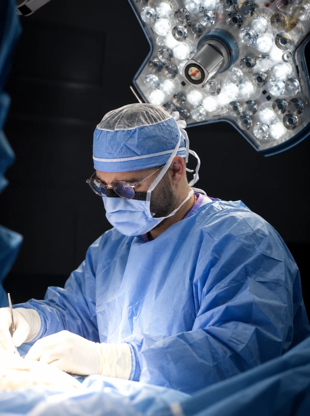
Reviewing surgeon
Foot and Ankle Surgeon at Limb Salvage Institute, a specialty practice of Ozturk Foot & Ankle. Dr. Ozturk is double board certified, as a Diplomate of the American Board of Podiatric Medicine (DABPM) and a Fellow of the Academy of Physicians in Wound Healing (FAPWH), and is a Fellow of the American Society of Podiatric Surgeons (FASPS) and the American Society of Podiatric Medicine (FASPM).
References
- 1.Bus SA, Sacco ICN, Monteiro-Soares M, et al. Guidelines on the prevention of foot ulcers in persons with diabetes (IWGDF 2023 update). Diabetes Metab Res Rev. 2024;40(3):e3651. PubMed 37302121 (external site)
- 2.Schaper NC, van Netten JJ, Apelqvist J, Bus SA, Fitridge R, Game F, et al. Practical guidelines on the prevention and management of diabetes-related foot disease (IWGDF 2023 update). Diabetes Metab Res Rev. 2024;40(3):e3657. PubMed 37243927 (external site)
- 3.Golledge J, Fernando ME, Alahakoon C, et al. Efficacy of at home monitoring of foot temperature for risk reduction of diabetes-related foot ulcer: A meta-analysis. Diabetes Metab Res Rev. 2022;38(6):e3549. PubMed 35605998 (external site)
- 4.Mueller MJ, Sinacore DR, Hastings MK, et al. Effect of Achilles tendon lengthening on neuropathic plantar ulcers. A randomized clinical trial. J Bone Joint Surg Am. 2003;85(8):1436-45. PubMed 12925622 (external site)
- 5.Murray HJ, Young MJ, Hollis S, Boulton AJ. The association between callus formation, high pressures and neuropathy in diabetic foot ulceration. Diabet Med. 1996;13(11):979-82. PubMed 8946157 (external site)
- 6.Margolis DJ, Allen-Taylor L, Hoffstad O, Berlin JA. Diabetic neuropathic foot ulcers: the association of wound size, wound duration, and wound grade on healing. Diabetes Care. 2002;25(10):1835-9. PubMed 12351487 (external site)
- 7.Young MJ, Cavanagh PR, Thomas G, Johnson MM, Murray H, Boulton AJ. The effect of callus removal on dynamic plantar foot pressures in diabetic patients. Diabet Med. 1992;9(1):55-7. PubMed 1551311 (external site)
- 8.Bus SA, Armstrong DG, Crews RT, et al. Guidelines on offloading foot ulcers in persons with diabetes (IWGDF 2023 update). Diabetes Metab Res Rev. 2024;40(3):e3647. PubMed 37226568 (external site)
- 9.Armstrong DG, Tan TW, Boulton AJM, et al. Diabetic Foot Ulcers: A Review. JAMA. 2023;330(1):62-75. PubMed 37395769 (external site)
- 10.Bonanno DR, Gillies EJ. Flexor Tenotomy Improves Healing and Prevention of Diabetes-Related Toe Ulcers: A Systematic Review. J Foot Ankle Surg. 2017;56(3):600-604. PubMed 28476394 (external site)
- 11.Andersen JA, Rasmussen A, Frimodt-Møller M, Kirketerp-Møller K, Rossing P. Long Term Outcomes After Flexor Tendon Tenotomy of the Diabetic Foot. Clin Med Insights Endocrinol Diabetes. 2025;18:11795514251314787. PubMed 39995589 (external site)
This article is general health information and isn’t a substitute for advice from a clinician who knows your history. Read our medical disclaimer and how we review content.
Learn more about this
Condition
Diabetic foot ulcers
Open sores on the feet of people with diabetes, and the step-by-step care, including surgery when it’s needed, that aims to heal them and keep them closed.
Treatment
Wound debridement
Removing callus and dead tissue from a foot wound, in the office or the operating room, so the wound can heal.
Treatment
Total contact casting
A knee-high cast or locked walker you can’t take off, which spreads pressure away from a foot ulcer so it can heal.
Treatment
Offloading surgery
Operations on the calf, Achilles or toe tendons (and sometimes a metatarsal bone) that take pressure off an ulcer so it heals and is less likely to come back.
Condition
Ulcers from foot deformity
Ulcers that form over bent toes, bunions and prominent bones on a foot with reduced feeling, and how offloading and surgery heal them.
Keep reading
Ulcers & offloading
Why a Foot Wound Deserves Prompt, Thorough Care
A painless sore on the foot can be more serious than it looks. What the research shows about waiting, what thorough wound care covers, and when surgery is the step that saves the foot.
8 min read
Limb salvage surgery
Achilles Lengthening for Forefoot Ulcers That Keep Coming Back
A tight calf can keep an ulcer under the ball of the foot from healing or staying healed. Who the operation suits, what the trials found and the trade-offs.
9 min read
Limb salvage surgery
Toe-Tip Ulcers and Flexor Tenotomy: A Small Procedure, a Big Benefit
An ulcer on the tip of a bent toe is usually a pressure problem with a small fix. How a flexor tenotomy works, who it suits, what the trials found and the trade-offs.
8 min read
On the Ozturk Foot & Ankle blog
General foot care and non-surgical basics, from our parent practice.