How to Diagnose Osteomyelitis in the Foot: Probe, X-ray, MRI and Bone Biopsy
Each test for foot bone infection points toward or away from the answer; a bone sample, by needle or during surgery, confirms it and names the germ. How each step works and what it can miss.
Medically reviewed by Dr. Efe Ozturk · Last reviewed
Published 7 min read
Hearing that a wound might have reached the bone is frightening. Getting the diagnosis right matters: bone infection greatly raises the risk of losing a toe, part of the foot or more, and an accurate answer avoids long courses of antibiotics, or an operation, that you may not need.[1] Here is how each test works, what it can miss, and why a bone sample is the step that gives a clear answer. For treatment options and outlook, see does osteomyelitis mean amputation?
Why is a bone biopsy the test that settles it?
Every other test points toward or away from the diagnosis. Only bone itself confirms it.[1,2]
- Wound swabs often name the wrong germ. In most studies, cultures of bone and of anything else agree less than half the time. In one direct comparison of 46 paired samples, a wound sample and a bone sample taken through the skin gave the same result in only 42%.[1]
- It can change the outcome. In a French study of 50 people treated without surgery, antibiotics chosen from a bone culture were the only factor linked with lasting remission: 18 of the 32 people in remission had received them, against 4 of the 18 who were not in remission.[3]
- It usually finds the germ. Across 11 studies of 837 needle biopsies, 84% grew bacteria in culture.[4]
How it is done. A needle biopsy passes through healthy, uninfected skin to avoid picking up germs from the wound. It is generally not painful, because most people with this problem have reduced feeling in the foot and a numbing injection can be given, and complications are rare.[1] Some experts suggest stopping antibiotics for a few days, ideally 2 weeks, before the biopsy to avoid a false-negative result, though the right gap is debated.[1]
Dr. Efe Ozturk, a Foot and Ankle Surgeon who is double board certified, including in wound care (FAPWH), performs bone biopsies when the result will guide treatment, and takes bone samples during surgery to remove infected bone. See surgery for bone infection.
What is the probe-to-bone test?
A sterile, blunt metal probe is gently slid into the wound. Touching hard, gritty bone is a positive test.[1] In a review of seven studies, it detected 87% of bone infections and correctly cleared 83% of wounds without one.[5] Along with a positive probe test, an ulcer larger than 2 square centimeters was one of the two most useful clinical clues.[2] The test depends on a skilled examiner, and two examiners don’t always agree.[1]
What does osteomyelitis look like on an X-ray?
X-rays come first for anyone with a possible bone infection, and we take them at the practice. Signs of infection include loss of the outer bone layer with erosion, thinning patches inside the bone, a lifted bone lining, hardened bone, and gas or abnormal tissue tracking down toward the bone. A fragment of dead bone sometimes shows.[1]
X-rays can miss early infection. Across 16 studies, they picked up 61.9% of bone infections.[6] So if the first film is normal and suspicion stays high, it is often repeated in 2 to 3 weeks.[1] An abnormal X-ray about doubles the odds of bone infection.[2]
Can blood tests show osteomyelitis?
A blood test for inflammation adds weight. An ESR (sedimentation rate) above 70 mm/h made bone infection much more likely in one review.[2] In a 2024 analysis of 19 studies, ESR, CRP and procalcitonin were all useful, but none was outstanding on its own.[7]
What does an MRI show?
MRI is the guideline’s strong recommendation when the diagnosis is still in doubt after the exam, X-rays and blood tests.[1] Across 22 studies, MRI detected 96.4% of bone infections and correctly cleared 83.8% of feet without one.[6] A normal MRI makes bone infection much less likely.[2]
MRI also maps how far the infection reaches and whether pus has collected, which helps a surgeon plan the operation.[1] We arrange MRI at an outside imaging center. When MRI isn’t possible, a PET scan or a white blood cell scan is an alternative.[1]
How do doctors tell osteomyelitis from Charcot foot?
Charcot foot is a breakdown of the foot’s bones and joints after nerve damage. Telling it apart from bone infection is a known challenge, and specialists advise relying on several findings rather than one test.[8] On X-ray, Charcot changes can look like infection, and on MRI, bone swelling from Charcot, an injury or earlier surgery makes the scan less specific.[1]
So the answer rests on several clues together: your history, the exam, X-rays and MRI, and blood tests. When infection is still suspected, a bone sample for culture and the microscope should be taken.[8] See Charcot foot and the Center for Lower Extremity Surgery’s page on Charcot foot (external site).
Where surgery fits once it is confirmed
The guideline suggests considering surgery to remove infected bone, with antibiotics, for foot bone infection, and antibiotics alone for a narrower group: a forefoot infection with no exposed bone, no artery disease and no need for urgent drainage.[1] In a randomized trial of forefoot bone infection under a nerve-related ulcer, 86.3% healed after bone-sparing surgery with 10 days of antibiotics, against 75% after 90 days of antibiotics alone, a difference that was not statistically significant; 4 people given antibiotics alone worsened and had surgery.[9] Surgery also shortens antibiotics: the guideline suggests up to 3 weeks after a minor amputation with infection left at the bone edge, against 6 weeks without surgery.[1] See bone infection of the foot.
Limits of these tests
No single test reliably rules bone infection in or out on its own.[1] Bone cultures can be falsely negative, especially after antibiotics, and no study has yet shown clearly that basing treatment on a biopsy improves outcomes, so the guideline’s biopsy advice is conditional.[1] Data on needle biopsy safety are limited.[4] And in the trial above, 3 people in the surgery group needed another operation.[9]
Common questions
Can an X-ray miss osteomyelitis in the foot?
Yes. X-rays missed more than a third of bone infections in pooled studies, especially early ones.[6] A normal film may be repeated in 2 to 3 weeks if suspicion remains.[1]
Is MRI or X-ray better for foot osteomyelitis?
MRI is more accurate, but X-rays come first because they are inexpensive and widely available and show other problems, such as gas or a foreign object.[1,6]
How do you differentiate osteomyelitis from Charcot foot?
By combining the history, exam, imaging and blood tests, and taking a bone sample when doubt remains.[8]
Which antibiotic works for foot osteomyelitis?
There is no single right choice. The guideline advises choosing based on the germ found and its sensitivities, how severe the infection is, and side effects.[1] That is why a bone culture matters.
The bottom line
Diagnosing osteomyelitis in the foot is a stepwise process: probe, X-ray, blood tests, then MRI if needed. The answer that names the germ comes from the bone itself, through a needle biopsy or during surgery. If a wound is deep, has lasted for weeks or keeps coming back, see a surgeon early, so the diagnosis and the operation, if one is needed, happen together.
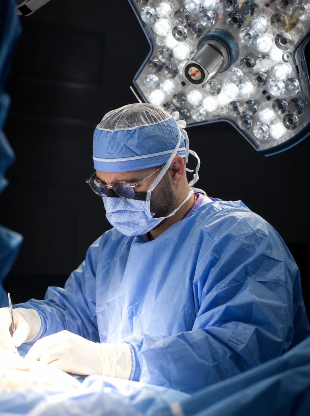
Reviewing surgeon
Foot and Ankle Surgeon at Limb Salvage Institute, a specialty practice of Ozturk Foot & Ankle. Dr. Ozturk is double board certified, as a Diplomate of the American Board of Podiatric Medicine (DABPM) and a Fellow of the Academy of Physicians in Wound Healing (FAPWH), and is a Fellow of the American Society of Podiatric Surgeons (FASPS) and the American Society of Podiatric Medicine (FASPM).
References
- 1.Senneville É, Albalawi Z, van Asten SA, Abbas ZG, Allison G, Aragón-Sánchez J, et al. IWGDF/IDSA guidelines on the diagnosis and treatment of diabetes-related foot infections (IWGDF/IDSA 2023). Diabetes Metab Res Rev. 2024;40(3):e3687. PubMed 37779323 (external site)
- 2.Butalia S, Palda VA, Sargeant RJ, Detsky AS, Mourad O. Does this patient with diabetes have osteomyelitis of the lower extremity? JAMA. 2008;299(7):806-13. PubMed 18285592 (external site)
- 3.Senneville E, Lombart A, Beltrand E, et al. Outcome of diabetic foot osteomyelitis treated nonsurgically: a retrospective cohort study. Diabetes Care. 2008;31(4):637-42. PubMed 18184898 (external site)
- 4.Schechter MC, Ali MK, Risk BB, et al. Percutaneous Bone Biopsy for Diabetic Foot Osteomyelitis: A Systematic Review and Meta-Analysis. Open Forum Infect Dis. 2020;7(10):ofaa393. PubMed 33134407 (external site)
- 5.Lam K, van Asten SA, Nguyen T, La Fontaine J, Lavery LA. Diagnostic Accuracy of Probe to Bone to Detect Osteomyelitis in the Diabetic Foot: A Systematic Review. Clin Infect Dis. 2016;63(7):944-8. PubMed 27369321 (external site)
- 6.Llewellyn A, Kraft J, Holton C, Harden M, Simmonds M. Imaging for detection of osteomyelitis in people with diabetic foot ulcers: A systematic review and meta-analysis. Eur J Radiol. 2020;131:109215. PubMed 32862106 (external site)
- 7.Ansert EA, Tarricone AN, Coye TL, et al. Update of biomarkers to diagnose diabetic foot osteomyelitis: A meta-analysis and systematic review. Wound Repair Regen. 2024;32(4):366-376. PubMed 38566503 (external site)
- 8.Rogero RG, Swamy S, Bettin CC. The Differentiation Between Infection and Acute Charcot. Orthop Clin North Am. 2024;55(2):299-309. PubMed 38403375 (external site)
- 9.Lázaro-Martínez JL, Aragón-Sánchez J, García-Morales E. Antibiotics versus conservative surgery for treating diabetic foot osteomyelitis: a randomized comparative trial. Diabetes Care. 2014;37(3):789-95. PubMed 24130347 (external site)
This article is general health information and isn’t a substitute for advice from a clinician who knows your history. Read our medical disclaimer and how we review content.
Learn more about this
Condition
Osteomyelitis (bone infection)
An infection of the bone in the foot, usually spreading from a diabetic foot ulcer, treated with culture-based antibiotics and often surgery to remove the infected bone.
Treatment
Osteomyelitis surgery
An operation to remove infected bone in the foot, usually beneath a diabetic foot ulcer, so the wound can heal, often with a shorter course of antibiotics.
Diagnosis guide
How wounds are evaluated
How a foot wound is evaluated, from the exam and measurements to the probe-to-bone test, cultures, X-rays, MRI, bone biopsy and circulation checks.
Condition
Charcot foot
Inflammation that weakens and collapses the bones of a numb foot, and the casting and surgery that keep the foot stable, ulcer-free and walking.
Condition
Diabetic foot infections
An infection of the skin, deeper tissue or bone of the foot in a person with diabetes, usually starting in a wound.
Keep reading
Infection & bone
Does Osteomyelitis Mean Amputation? Bone Infection in the Foot
A bone infection in the foot is serious, but most people keep their leg. How it is treated, what the studies show, and when surgery is the step that saves the foot.
9 min read
Limb salvage surgery
Can a Collapsed Charcot Foot Be Rebuilt? What Reconstruction Achieves
A collapsed Charcot foot can often be realigned and fused so it rests flat again. What reconstruction achieves, who it suits and what it asks of you.
8 min read
Ulcers & offloading
Why Diabetic Foot Ulcers Don’t Heal: The Five Usual Reasons and What Fixes Each
A stalled ulcer usually has a cause a dressing can’t reach, and four of the five usual causes have a surgical or procedural answer. How each is found, what fixes it, and when surgery closes the wound.
7 min read
On the Ozturk Foot & Ankle blog
General foot care and non-surgical basics, from our parent practice.