Tenex for plantar fasciitis: how the procedure works, results, recovery and risks
A deep look at the Tenex procedure for plantar fasciitis: how the ultrasonic probe works, who is a candidate, the published heel studies, and what recovery looks like week by week.

By Dr. Efe Ozturk
· 10 min read
Medically reviewed by Dr. Efe Ozturk · Last reviewed
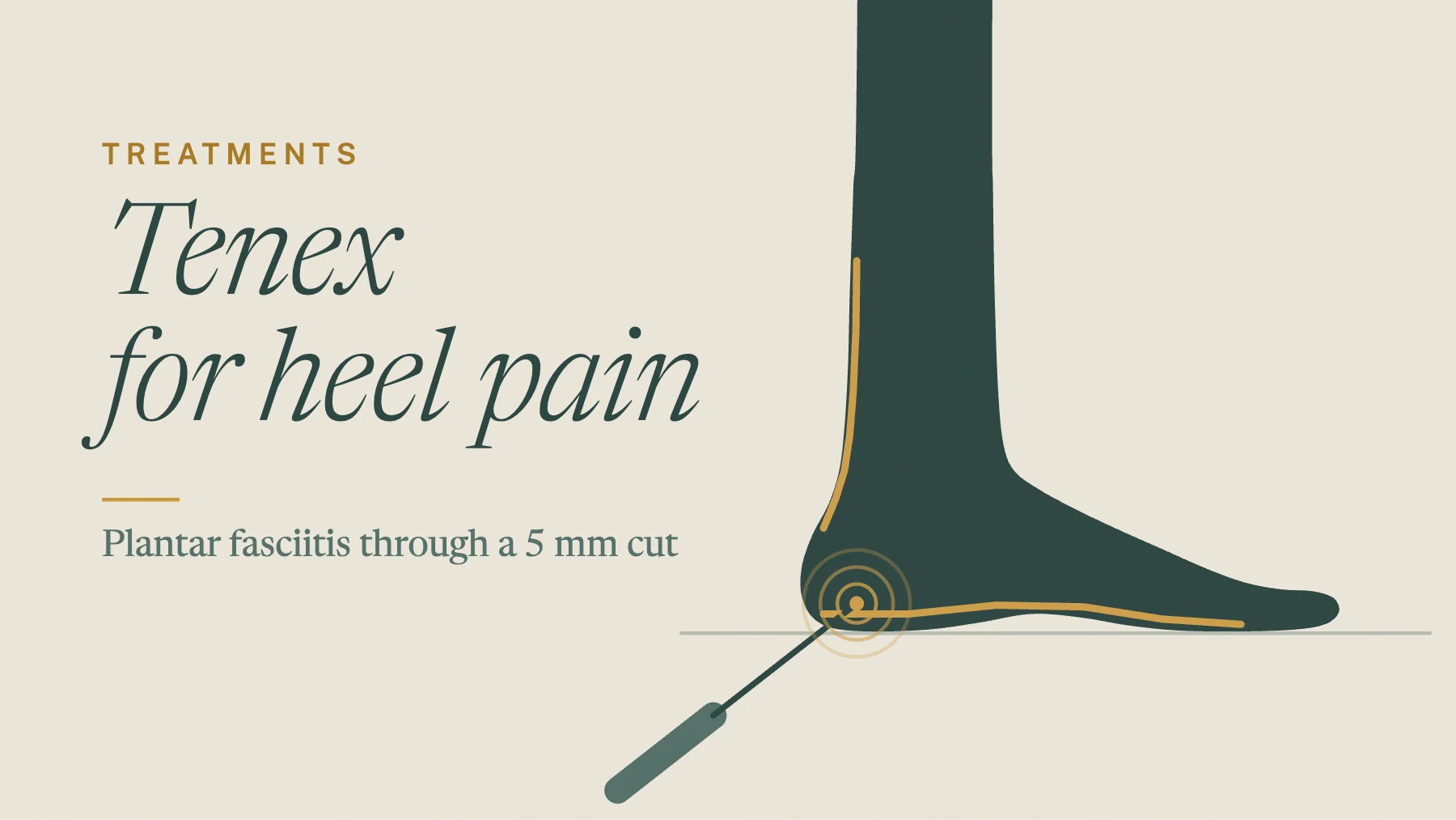
If you’ve had heel pain for six months or more, you’ve probably heard the word Tenex. This guide covers one device for one problem: what the Tenex procedure for plantar fasciitis does, who it suits, what the day and the weeks after look like, and what the published heel studies found. For the side-by-side with TenJet, see our Tenex and TenJet overview.
Why consider a procedure instead of waiting?
Plantar fasciitis can last for years. In a Danish study of 174 people, half still had it five years after it began, and those who did recover had pain for about two years first.[6] That’s a long time to limp through mornings.
The case for acting comes from randomized trials. In one, endoscopic surgery plus strength training gave better foot function at one year than cortisone shots plus the same training, and more people returned to running and jumping.[7] In another, for people with tight calves, calf lengthening plus stretching left pain at 2.5 out of 10 at six years, versus 5.5 with stretching alone.[8] Neither trial tested Tenex. They do show that, for the right heel, a procedure can do what more months of the same care did not.
Tenex is the smallest step on that surgical ladder. It goes straight to the damaged tissue you can see on ultrasound, without opening the heel.
What is the Tenex procedure for plantar fasciitis?
Tenex is also called percutaneous ultrasonic tenotomy or fasciotomy. “Percutaneous” means through the skin. “Fasciotomy” means cutting into the fascia, the tough band under your arch.
- The tool. The TX1 and TX2 handpieces work the same way. The tip vibrates with ultrasonic energy, which cuts and breaks up damaged tissue while saline rinses the area and suction carries the debris away.[1,9]
- The size. The probe is about as thin as an 18-gauge needle.[9]
- The target. In plantar fasciitis, the fascia near the heel looks thickened on ultrasound.[6] The probe releases the diseased tissue where it attaches to the heel bone.[9] The device was designed to remove only diseased tissue.[10]
- The clearance. The FDA cleared the Tenex Health TX System in 2016 as an ultrasonic surgical instrument.[5]
In an early study of the device, at the elbow, the procedure itself took a median of about 10 minutes, with the energy switched on for about half a minute of that.[10]
Who is a candidate for Tenex?
You may be a good candidate if:
- Heel pain has lasted about six months despite good care. That’s when the American College of Foot and Ankle Surgeons says surgery can be considered.[2] Good care means stretching, supportive shoes or inserts, and often shockwave. Our plantar fasciitis treatment guide covers those first steps.
- Ultrasound shows a thickened, damaged area at the heel that a probe can reach.
- A release didn’t work. In the first Tenex heel series, four of the 12 patients had already had an open or endoscopic release.[9]
Some heels need a different plan. A tight calf may do better with calf lengthening.[8] Burning or tingling points to a nerve such as Baxter’s nerve entrapment (external site). And a heel spur on X-ray isn’t, by itself, a reason for Tenex. OrthoInfo notes that spurs do not cause plantar fasciitis pain, and most people with spurs have no heel pain.[11] See heel spurs and heel pain.
What is the Tenex plantar fasciitis success rate?
Here are the published heel series we found, with the device named where the abstract names it.
| Study | Patients | Follow-up | What happened |
|---|---|---|---|
| Ha 2025, Mayo Clinic[3] | 58 procedures | 6.6 years | Pain 6.8 to 0.7; first-step pain 0.9; 91% successful; 90% would recommend it; no complications |
| Kruse 2024[12] | 67 | 1 year | Ultrasonic fasciotomy; pain improved 5.87 points; 94% satisfied at 12 and 52 weeks; no complications |
| Encinas 2025[4] | 56 (Tenex) | 27 months | PROMIS pain improved 5.2 points, with gains in function and mobility |
| Patel 2015[9] | 12 (Tenex) | 2 years | Foot score 30 to 88 after surgery; all 12 essentially pain-free at 2 years |
| Fick 2021[1] | 80 (Tenex) | 6 weeks to several years | 36 of 57 who answered were satisfied early; physical function scores rose from 36 to 48 |
| Turner 2024[13] | 30 | About 10 months | Ultrasonic fasciotomy; pain fell with or without added PRP, more with PRP |
Two points stand out. People in these studies had long-standing pain, about two years on average in the Mayo series.[3] And the relief held: the Mayo results were measured more than six years out.
A 2023 meta-analysis pooled 35 studies of ultrasound-guided needle tenotomy, a group of techniques that includes Tenex, in tendons and fascia across the body. Pain and function improved in the short, middle and long term. The largest gains came in the first three months, and different tenotomy methods did about the same.[14]
Does an add-on injection help? In the Turner series, people who also got platelet-rich plasma (PRP, a concentrate of your own blood) had a larger drop in pain.[13] In another small study, an amniotic tissue injection added to ultrasound-guided fasciotomy helped pain early, but the two groups were the same at one year.[15]
What happens on the day of a Tenex procedure?
- Mapping. Ultrasound finds the damaged fascia and the nearby nerves, the medial calcaneal and lateral plantar nerves, before any cut.[1]
- Numbing. About 5 to 10 mL of local anesthetic numbs the heel. In the Iowa series, no one needed sedation.[1]
- The cut. A small blade makes a cut of about 5 mm on the inner side of the heel, in line with the fascia fibers.[1,9]
- Treatment. Watching on the ultrasound screen, the surgeon moves the probe through the damaged area until it has all been treated.[9]
- Closing. A stitch and adhesive strips and a compressive dressing go on, and you leave in a walking boot.[9]
You walk out the same day.
What is the Tenex plantar fasciitis recovery time?
This timeline follows the published Iowa rehabilitation plan for the foot and ankle and the first Tenex heel series. Your own plan depends on how much tissue was treated.
- Days 1 to 3. Ice and simple pain relievers as needed. The dressing comes off after two to three days.[9]
- Week 1. Walk as comfortable in the boot. Gentle range of motion starts the day after. No barefoot walking.[1]
- Weeks 1 to 2. Stop the boot once you can walk without pain or a limp. The stitch and strips come out at 10 to 14 days.[1,9]
- Weeks 2 to 6. Gentle strengthening and balance work. No running, jumping, cutting or pivoting yet.[1]
- Weeks 5 to 13. This is when pain eased in the first Tenex heel series.[9]
- Week 6 onward. Load builds step by step. Six weeks was the earliest anyone returned to full activity in the Iowa series.[1]
- Week 12 onward. Most people going back to sport or manual work needed about 12 weeks before full clearance.[1]
How does Tenex compare with plantar fascia release surgery?
Plantar fascia release works. In a review of 26 endoscopic studies covering 978 feet, foot scores rose from about 56 to 90 out of 100.[16] But a release partly cuts the fascia where it attaches to the heel,[11] and its published complication rates are higher:
| Approach | Published complication rate |
|---|---|
| Open release | 11%[17] |
| Endoscopic release | 5% in one review, 8.9% in another[17,16] |
| Ultrasonic tenotomy, Tenex | 0.7% across 289 procedures at several body sites; none reported in the Mayo or Kruse heel series[1,3,12] |
These numbers come from different patients, not a head-to-head trial, so they show the general picture only. Still, the appeal is clear: a 5 mm cut, local numbing, and walking the same day. If the damaged area can be targeted, we offer Tenex before an open release. Open and endoscopic surgery are done through our Center for Lower Extremity Surgery (external site).
How does Tenex compare with shockwave, PRP and cortisone?
No study has compared Tenex directly with any of them for heel pain. Here is what each has shown on its own:
- Shockwave. In a review of 236 trials, shockwave was the only treatment that improved pain in the medium and long term compared with control.[18] We offer shockwave therapy, and it is a reasonable step before a procedure.
- PRP. The same review found PRP helped pain in the short term.[18]
- Cortisone. A Cochrane review found a shot may slightly ease heel pain for up to a month, but not after that. Two plantar fascia ruptures were reported among 699 people given steroid shots.[19]
In our view, the difference is in kind. Injections and shockwave aim to change how the tissue heals, while Tenex removes the damaged tissue itself.
What are the risks of Tenex for plantar fasciitis?
In the largest safety series, 2 of 289 procedures had a complication: one superficial wound infection and one area of skin sensitivity.[1] No complications were reported in the Mayo or Kruse heel series, and the first heel series had no infections or wound problems.[3,12,9] Nerves run close to the heel, so they are mapped on ultrasound before the cut.[1] And it doesn’t work for everyone: in the Mayo series, 5 of 58 procedures were not rated a success.[3]
What are the limits of the evidence?
We found no randomized trial of Tenex for plantar fasciitis. None of the heel studies above compares Tenex with another treatment, and a 2025 systematic review of needle tenotomy and Tenex graded its evidence at level 4, the level of the weakest studies it included.[20] The Mayo and Kruse abstracts describe the technique but don’t name the device.[3,12] In 2018, the American College of Foot and Ankle Surgeons rated ultrasonic debridement “uncertain,” before most of these results were published.[2] A 2026 network review of 21 surgical studies found endoscopic approaches improved scores more than open or percutaneous techniques, and calf lengthening gave better pain scores, though the studies had moderate to high risk of bias.[21] The first Tenex heel study’s author sat on the company’s medical advisory board, and an author of the Iowa series consulted for it.[9,1]
How we decide
Dr. Efe Ozturk is a Foot and Ankle Surgeon and podiatrist who offers both Tenex and TenJet. We confirm the diagnosis with an exam and in-office ultrasound, check your calf and nerves, and make sure the first steps had a fair trial. Then we lay out the options side by side: Tenex, TenJet, calf lengthening or a release. See our heel pain page. We see patients in Lyndhurst, Paramus and Millburn, NJ. Please call our office at 908-215-5953 to inquire about your insurance.
Common questions
Does the Tenex procedure hurt?
The heel is numbed first, and in the Iowa series no one needed sedation.[1] Afterward, ice and simple pain relievers were enough for the first few days in the first Tenex heel series.[9]
How long does the Tenex procedure take?
At the elbow, the procedure itself took a median of about 10 minutes.[10] Plan on a longer visit for mapping, numbing and the dressing.
Can Tenex remove a heel spur?
For plantar fasciitis, Tenex treats the damaged fascia, not the spur. Spurs do not cause plantar fasciitis pain, and in the Danish study a spur made no difference to how people recovered.[11,6]
Is Tenex better than shockwave or PRP for plantar fasciitis?
That hasn’t been tested. In a large review, shockwave was the only non-surgical treatment that helped pain in the medium and long term.[18] Tenex is the option that removes damaged tissue when those haven’t worked.
When to see a Foot and Ankle Surgeon or podiatrist
About the author
Dr. Efe Ozturk
Foot and Ankle Surgeon. Dr. Ozturk is double board certified, as a Diplomate of the American Board of Podiatric Medicine (DABPM) and a Fellow of the Academy of Physicians in Wound Healing (FAPWH), and is a Fellow of the American Society of Podiatric Surgeons (FASPS) and the American Society of Podiatric Medicine (FASPM). Trained in forefoot and rearfoot reconstructive surgery at Morristown Medical Center; sees patients in Lyndhurst, Paramus and Millburn.
Where to be seen
Dr. Ozturk sees patients at each of these offices, and one phone number reaches all of them.
References
- 1.Fick B, Stover DW, Chimenti RL, Hall MM. The Safety of Ultrasound Guided Tenotomy and Debridement for Upper and Lower Extremity Tendinopathies: A Retrospective Study. Iowa Orthop J. 2021;41(2):82-90. PubMed 34924874 (external site)
- 2.Schneider HP, Baca JM, Carpenter BB, Dayton PD, Fleischer AE, Sachs BD. American College of Foot and Ankle Surgeons Clinical Consensus Statement: Diagnosis and Treatment of Adult Acquired Infracalcaneal Heel Pain. J Foot Ankle Surg. 2018;57(2):370-381. PubMed 29284574 (external site)
- 3.Ha CT, Romero JM, Gonzalez-Carta K, et al. Ultrasound-Guided Percutaneous Fasciotomy and Debridement for Chronic Plantar Fasciopathy: Six-Year Patient-Reported Outcomes from a Retrospective Case Series. Foot Ankle Int. 2025;46(11):1239-1247. PubMed 41178389 (external site)
- 4.Encinas R, Hall S, Gauthier C, et al. Minimally invasive ultrasound-guided percutaneous plantar fasciotomy on chronic plantar fasciitis: A retrospective analysis. Foot Ankle Surg. 2025;31(8):737-741. PubMed 40399170 (external site)
- 5.U.S. Food and Drug Administration. 510(k) Premarket Notification K153299: Tenex Health TX System. Decision date March 3, 2016. accessdata.fda.gov (external site)
- 6.Hansen L, Krogh TP, Ellingsen T, Bolvig L, Fredberg U. Long-Term Prognosis of Plantar Fasciitis: A 5- to 15-Year Follow-up Study of 174 Patients With Ultrasound Examination. Orthop J Sports Med. 2018;6(3):2325967118757983. PubMed 29536022 (external site)
- 7.Johannsen F, Konradsen L, Herzog R, Krogsgaard MR. Endoscopic fasciotomy for plantar fasciitis provides superior results when compared to a controlled non-operative treatment protocol: a randomized controlled trial. Knee Surg Sports Traumatol Arthrosc. 2020;28(10):3301-3308. PubMed 32006073 (external site)
- 8.Riiser MO, Husebye EE, Hellesnes J, Molund M. Outcomes After Proximal Medial Gastrocnemius Recession and Stretching vs Stretching as Treatment of Chronic Plantar Fasciitis at 6-Year Follow-up. Foot Ankle Int. 2024;45(1):1-9. PubMed 37902240 (external site)
- 9.Patel MM. A novel treatment for refractory plantar fasciitis. Am J Orthop (Belle Mead NJ). 2015;44(3):107-110. PubMed 25750942 (external site)
- 10.Koh JS, Mohan PC, Howe TS, et al. Fasciotomy and surgical tenotomy for recalcitrant lateral elbow tendinopathy: early clinical experience with a novel device for minimally invasive percutaneous microresection. Am J Sports Med. 2013;41(3):636-644. PubMed 23302261 (external site)
- 11.American Academy of Orthopaedic Surgeons. Plantar Fasciitis and Bone Spurs. OrthoInfo. orthoinfo.org (external site)
- 12.Kruse RC, Volfson E. Ultrasonic Fasciotomy for the Treatment of Chronic Plantar Fasciopathy: A Prospective Study. Clin J Sport Med. 2024;34(4):335-340. PubMed 38407211 (external site)
- 13.Turner A, Wang J, Liu G, Wukich D, VanPelt M. Retrospective Evaluation of Ultrasound Guided Percutaneous Plantar Fasciotomy With and Without Platelet Rich Plasma. J Foot Ankle Surg. 2024;63(2):233-236. PubMed 38043602 (external site)
- 14.Shomal Zadeh F, Shafiei M, Shomalzadeh M, Pierce J, Thurlow PC, Chalian M. Percutaneous ultrasound-guided needle tenotomy for treatment of chronic tendinopathy and fasciopathy: a meta-analysis. Eur Radiol. 2023;33(10):7303-7320. PubMed 37148349 (external site)
- 15.Nakagawa H, Sung K, Ashkani-Esfahani S, Waryasz G, May T, Sussman WI. Plantar fasciitis: a comparison of ultrasound-guided fasciotomy with or without amniotic membrane allograft injection. Regen Med. 2022;17(12):931-940. PubMed 36222008 (external site)
- 16.Ward L, Mercer NP, Azam MT, et al. Outcomes of Endoscopic Treatment for Plantar Fasciitis: A Systematic Review. Foot Ankle Spec. 2025;18(2):193-201. PubMed 36342049 (external site)
- 17.Saghir MA, Bashandy AS, Kholeif AM. Endoscopic plantar fasciotomy versus open plantar fasciotomy for treatment of plantar fasciitis: a systematic review and meta-analysis. J Orthop Surg Res. 2026;21(1):449. PubMed 42204579 (external site)
- 18.Guimarães JS, Arcanjo FL, Leporace G, et al. Effects of therapeutic interventions on pain due to plantar fasciitis: A systematic review and meta-analysis. Clin Rehabil. 2023;37(6):727-746. PubMed 36571559 (external site)
- 19.David JA, Sankarapandian V, Christopher PR, Chatterjee A, Macaden AS. Injected corticosteroids for treating plantar heel pain in adults. Cochrane Database Syst Rev. 2017;6(6):CD009348. PubMed 28602048 (external site)
- 20.Maag L, Linder S, Hackett L, et al. Effectiveness of Percutaneous Needle Tenotomy for Tendinopathies: A Systematic Review. Sports Health. 2025;17(4):834-842. PubMed 39238190 (external site)
- 21.Tan AKS, Lee DMX, Mai AS, Toh RX, Li ZX, Mohamed Buhary KS, et al. Comparative effectiveness of surgical interventions for recalcitrant plantar fasciitis: A systematic review and network meta-analysis. J Foot Ankle Surg. 2026. PubMed 42604712 (external site)
This is general health information and isn’t a substitute for advice from a clinician who knows your history. Read our medical disclaimer and how we write and review content.
More from the blog
Treatments
TenJet for plantar fasciitis: how the procedure works, what the evidence shows, and recovery
How the TenJet saline jet treats stubborn plantar fasciitis through a small incision, who it suits, what the evidence does and doesn’t show yet, and recovery week by week.
Treatments
TenJet for Achilles tendonitis: how the procedure works, who it suits, and recovery
How the TenJet saline jet treats stubborn Achilles tendonitis through a small incision, how it differs from Tenex, what the evidence does and doesn’t show, and recovery week by week.
Treatments
Tenex for Achilles tendonitis: insertional vs midportion, Haglund’s, results and recovery
A deep look at the Tenex procedure for the Achilles: results at the heel bone and higher up, why a Haglund’s bump or calcium matters, how it compares with open surgery, and recovery week by week.